Chest anatomy, 19th Century illustration

![]()
Wall Art and Photo Gifts from Science Photo Library
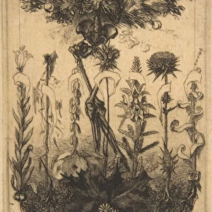
Chest anatomy, 19th Century illustration
Chest anatomy, 19th Century illustration. Historical hand coloured lithographic print showing the lungs (centre) in the chest, larynx (voicebox, white, upper centre) in the throat, and the blood vessels (red and blue) and muscles (brown) of the neck. The lungs are protected by the ribs, which are connected by the intercostal muscles. Image from Traite complet de l anatomie de l homme, comprenant la medecine operatoire Vol. 4 (1836), by Jean-Baptiste Marc Bourgery and illustrated by Nicolas-Henri Jacob
Science Photo Library features Science and Medical images including photos and illustrations
Media ID 6327677
© SCIENCE PHOTO LIBRARY
1836 Bones Carotid Chest Circulatory Descriptive Anatomy Diagram Diaphragm French Frontal Interior Internal Larynx Lithograph Lithographic Print Lung Lungs Muscles Neck Organs Plate 1 Pulmonary System Respiratory Ribs Shoulder Shoulders System Thoracic Thorax Throat Upper Arm Vascular System Vessels Vol 4 Volume Four Circulation
EDITORS COMMENTS
This 19th-century lithographic print showcases a detailed illustration of chest anatomy. The historical hand-colored image provides a comprehensive view of the intricate structures within the human chest. At its center, the lungs take prominence, surrounded by an array of blood vessels and muscles that form the neck. The ribs, connected by intercostal muscles, serve as protective barriers for these vital organs. Originally featured in Jean-Baptiste Marc Bourgery's renowned medical treatise "Traite complet de l'anatomie de l'homme" this artwork was skillfully illustrated by Nicolas-Henri Jacob. Its artistic depiction beautifully captures the complexity and beauty of the human body. The print offers valuable insights into various anatomical elements such as the throat (housing the larynx or voicebox), blood circulation (represented through red and blue vessels), and muscular structure (depicted in brown). It serves as a visual guide to understanding how these components work together harmoniously. With a white background highlighting every detail, this lithograph is not only an exquisite piece of art but also an invaluable resource for scientific study. Its inclusion in Volume Four of Bourgery's treatise demonstrates its significance in advancing medical knowledge during that era. This remarkable print from Science Photo Library allows us to appreciate both the aesthetic appeal and educational value inherent in historical anatomical illustrations like this one.
MADE IN THE USA
Safe Shipping with 30 Day Money Back Guarantee
FREE PERSONALISATION*
We are proud to offer a range of customisation features including Personalised Captions, Color Filters and Picture Zoom Tools
SECURE PAYMENTS
We happily accept a wide range of payment options so you can pay for the things you need in the way that is most convenient for you
* Options may vary by product and licensing agreement. Zoomed Pictures can be adjusted in the Cart.


![[Young Woman, Nude, Full Figure in Profile], 1860s. Creator: Unknown](/sq/731/young-woman-nude-figure-profile-1860s-20170442.jpg.webp)